000
1. What is SPECT-CT?
Single Photon Emission and X-ray Computed Tomography System (Fig.1), for short SPECT-CT, is a combined system, which can obtain SPECT images with metabolic function information and CT images with anatomical structure information in once imaging (Fig.2).
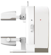
Fig.1. The model machine of SPECT-CT
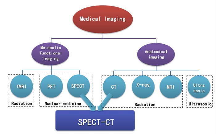
Fig.2. Medical imaging
Explanatory note: fMRI (functional Magnetic Resonance Imaging), PET (Positron Emission Tomography), SPECT (Single Photon Emission Computed Tomography), CT (Computer Tomography), MRI (Magnetic Resonance Imaging).
Traditional SPECT Imaging
SPECT imaging detects and records gamma rays emitted by radioactive tracers introduced into target tissues or organs in human body, and displays them in the form of images (Fig.3). It can provide information about blood flow, function, metabolism and receptors of organs or lesions, which can realize early diagnosis of diseases. However, it cannot reflect detailed anatomical structure information and achieve precise location of lesions.
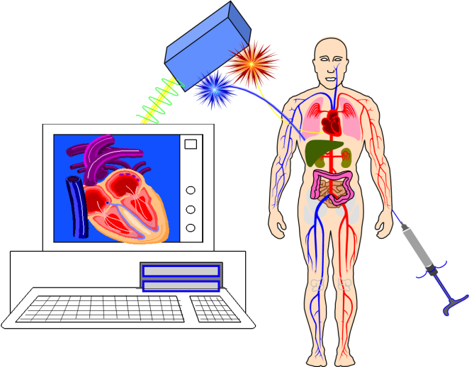
Fig.3. SPECT imaging schematic
Traditional CT Imaging
CT imaging uses external X-rays to rotate around the layer of a certain thickness of the human body examination areas, and receives x-rays through the layer, which are converted by the calculation program and displayed in the form of images (Fig.4). It can provide structural information and accurate anatomical location of organs or lesions. However, it cannot reflect functional status.

Fig.4. CT imaging schematic
(Source: http://slidesplayer.com)
SPECT-CT Imaging
SPECT-CT imaging through the sequential execution of SPECT and CT imaging (or CT imaging before SPECT imaging) in a single scan procedure to obtain both SPECT and CT images at one time. And then multimodal image fusion technology is used to give full play to the advantages of SPECT and CT, and make up for their shortcomings, improve the image information, and achieve 1+1>2 diagnostic efficiency. In addition, CT images can be used to accurately correct the attenuation of SPECT images, which provides a reliable basis for quantitative analysis of SPECT images.
1. The technical advantages of SPECT-CT
SPECT-CT fusion image
SPECT-CT fusion image is computed using multimodal image fusion technique, compared with single mode SPECT image, the SPECT-CT fusion image can provide valuable anatomical information for the correct interpretation of SPECT image, and can clearly locate the lesion and provide tissue structure information around the lesion. It can significantly improve the accuracy of nuclear medicine diagnosis. (Fig.5 and Fig.6 are examples of fusion images of SPECT-CT.)
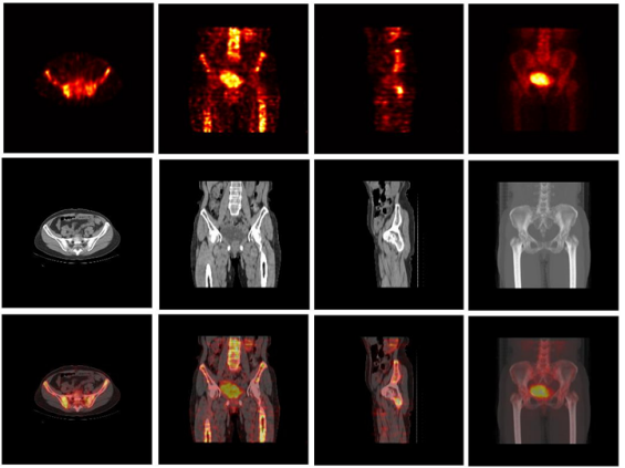
Fig.5. SPECT-CT images of the pelvic region. (The three rows from top to bottom are SPECT images, CT images and SPECT-CT fusion images. The four columns from left to right are the cross-section, coronal section, sagittal section and 3D MIP image of the scanning site.)
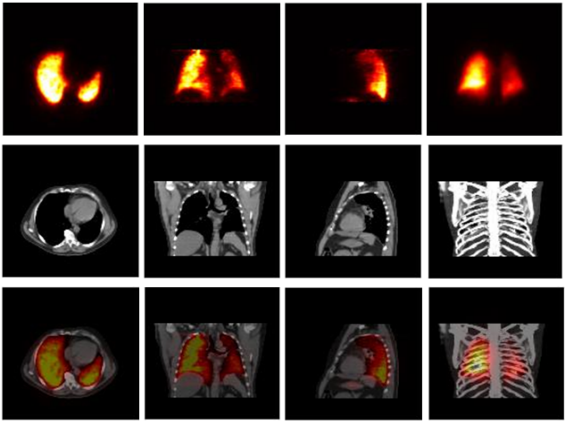
Fig.6. SPECT-CT images of the chest region. (The three rows from top to bottom are SPECT images, CT images and SPECT-CT fusion images. The four columns from left to right are the cross-section, coronal section, sagittal section and 3D MIP image of the scanning site.)
Accurate attenuation correction for SPECT image
Due to the attenuation of gamma ray in human body, the functional parameters measured in clinical for SPECT image often have some errors with the real value, which makes the accurate quantitative calculation of SPECT impossible. And the attenuation of gamma rays in the human body is nonlinear uneven, it is closely related to the density and thickness, therefore, the organizational structure to carry information of CT image became the best model of attenuation of gamma correction, so as to make the quantitative calculation of SPECT image possible (FIig.7 and Fig.8, respectively is uniform water model and SPECT images of pulmonary ventilation).

Fig.7. SPECT reconstruction images of uniform water model. (a) CT image of water model, (b) reconstruction result image of filtered back projection method (FBP), (c) reconstruction result image of iterative method (OSEM), (d) reconstruction result image with CT attenuation correction. (In images (b) and (c), the edge is bright and the middle is dark, which is caused by the nonlinear and non-uniform attenuation of γ ray itself in the tissue. Image (d) shows that attenuation correction using CT has effectively solved the problem of gamma ray attenuation.)
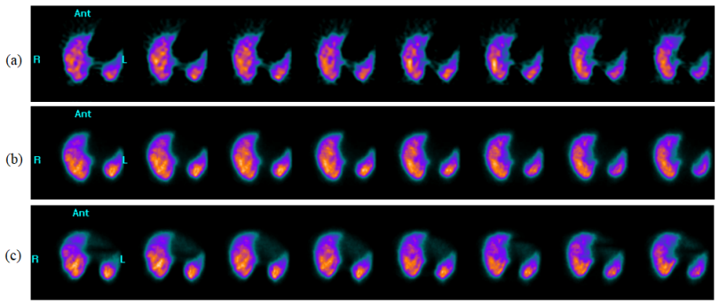
Fig.8. SPECT images of lung ventilation. (a) is the result of FBP reconstruction, (b) is the result of OSEM reconstruction, and (c) is the result of OSEM reconstruction with CT attenuation correction.
In combination with the technical advantages of SPECT-CT, we can say that the appears of SPECT-CT has transformed nuclear medicine from 'Unclear Image'!
(Unclear Image)!
1. Clinical application and market prospect of SPECT-CT
As the combination of SPECT system and CT system, SPECT-CT can not only realize the combined examination of osteology, cardiology, oncology and neurology, but also can perform separate nuclear medicine examination or radiology examination, with all the functions of nuclear medicine equipment and CT equipment. It can meet the clinical application requirements of nuclear medicine and radiology.
With its advantages in technology and clinical application, the market demand for SPECT-CT is increasing year by year. By the end of 2019, there were 903 single-photon imaging devices in China (Fig.9). SPECT-CT accounted for 55% of the 903 single-photon imaging devices, with an increasing trend year by year. From the current development trend, the domestic market and clinical application of single photon imaging equipment tend to SPECT-CT, single SPECT is gradually marginalized.
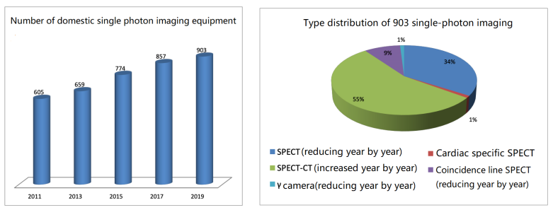
Fig.9. Statistics on the number and type distribution of domestic single photon imaging equipment from 2011 to 2019.
(Source of statistical data: Bulletin on the Current Situation of Nuclear Medicine in China, Chinese Society of Nuclear Medicine.)
What is more noteworthy is that domestic SPECT-CT has no registered products so far, SPECT-CT can only rely on imports, about 50 per year, and the annual growth rate of about 30%. Therefore, the future development of China's nuclear medical imaging equipment needs the joint efforts of domestic medical imaging equipment manufacturers and nuclear medicine practitioners to realize the clinical use of domestic SPECT-CT as soon as possible and break the foreign monopoly.
Hamamatsu Photon Technology (Langfang), a wholly-owned subsidiary of Beijing Hamamatsu Photon Techniques INC., has been deeply engaged in the development of nuclear medicine imaging equipment for 22 years, and has always been committed to the research of system and application. It can provide complete machine, components and other services for medical institutions and enterprises engaged in the field of nuclear medicine, and jointly incubate SPECT and SPECT-CT products. To provide rich soil for the development of domestic nuclear medicine infrastructure equipment, to promote the development of medical cause, to serve the progress of human health.